


An article authored by Dr Liron Levy-Hirsch DVM MRCVS
Liron graduated from the Hebrew University in Israel in 2007. After completing a one-year small animal rotating internship at the Hebrew University Veterinary Hospital, he moved to the UK to advance his career in veterinary medicine. Following experience in general practice, Liron joined Vets-Now Ltd., where he practiced Emergency and Critical Care in both clinical and leadership roles for over 12 years. In 2023, Liron joined HT VET as Managing Director.
Lumps and bumps are common findings in dogs. As veterinary professionals we see a range of worry from owners, from the deeply concerned to the “I’m sure it’s nothing.” The truth is, without a proper diagnostic workup, it is impossible to know the true nature of a lump, and our role as advocates for animal health is to ensure each lump is given the time and respect to determine what it is. There are numerous options available to practitioners when it comes to assessing dermal and subcutaneous masses, and this article will explore common diagnostic methods, the limitations of certain techniques, new advancements in veterinary technology, and how artificial intelligence (AI) is transforming cancer screening.
When a lump or bump is noticed on a dog, the first step is to locate it and asses if it is dermal, subcutaneous or within deeper tissues. Next is understanding their nature—whether they are benign or malignant, infectious, inflammatory, or caused by trauma. While many lumps are harmless, early identification and diagnosis of potentially malignant masses are critical for improving outcomes.
At the initial consultation for a lump or bump, the most important thing to do, is something! The “pea for a month” saying – if a lump is pea sized or bigger and has been there for a month or more – should be followed and all lumps and bumps in this category should be investigated. This begins with palpation, to assess size, shape, consistency, mobility and whether it is painful to the touch. However, palpation alone shouldn’t be relied upon when diagnosing. Further investigations such as an FNA or biopsy, particularly if done early and before excision, can lead to improved patient outcomes.
New screening tool

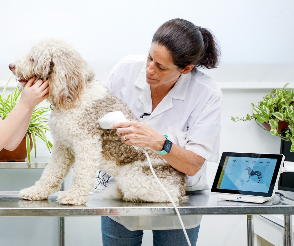
However, there are situations when gaining permission to move straight to invasive diagnostic testing is met with resistance from pet owners. Or times where the clinician feels the lump is benign but would prefer more information to confirm this assumption. A new screening tool, the HT Vista, helps to bridge this gap and offers on the spot screening of masses. The HT Vista uses Heat Diffusion Imaging (HDI) technology to record the tissue’s thermogenic behaviour as it gets heated and then cools down naturally. This information is analysed using an AI-based algorithm which then provides a score from 1-10. A high score of 5-10 indicates the mass is likely to be benign at a high probability between 90% and 99%, whereas a low score of 1-4 indicates a lower probability of the mass being benign and, therefore, recommends further diagnostic testing.
The use of a screening device ensures more masses are scanned and screened, which typically results in more masses being aspirated or biopsied, which is essential when trying to catch cases of cancer early.
Obtaining a definitive diagnosis with cytology or biopsy early results in early implementation of treatment plans. The earlier a mass is detected, the more likely surgery will be curative, particularly for small tumours. If a tumour is removed with complete surgical margins, prognosis is often favourable.
Identifying a mass

Before moving to surgery, it is important to know what the mass is before you remove it. For the majority of cases this can be achieved with an FNA and cytology. FNA can help to distinguish neoplasia, such as mast cell tumours and soft tissue sarcomas, from inflammation and benign masses, including lipomas and sebaceous adenomas. For malignant tumours, cytology provides information that assists in formulating diagnostic and treatment plans. While FNA is relatively quick and affordable to obtain, it is important to remember that due to the small number of cells obtained, it may not provide enough information to fully characterise a tumour’s behaviour and it is not always diagnostic.
If an FNA is non-diagnostic, a biopsy and histopathology is recommended before surgical excision. Depending on the size of the mass either a punch biopsy or incisional biopsy is required. A biopsy provides a more comprehensive understanding of the mass, offering a definitive diagnosis. This will help determine the best treatment plan leading towards a better prognosis.
While it is tempting to provide excisional biopsies, this should be avoided if possible. It is far better to surgically remove a mass once its nature has been determined, to ensure adequate margins have been taken. Incomplete margins result in more treatment and a poorer prognosis.
Treatment plan
Once a diagnosis and staging have been made, a treatment plan can commence. In many cases, surgical excision itself is enough, particularly if it has been performed as early as possible. Some of the more common malignant masses we see in dogs are mast cell tumours, soft tissue sarcomas, and squamous cell carcinomas. It is imperative that these masses are not missed at the initial consultation with the “wait and see” method. It is also important to understand that these masses can often look and feel like a lipoma, but each lump and bump should be appropriately screened, and offered further diagnostics if necessary.
Assessing lumps and bumps in dogs is a vital part of veterinary care, requiring a combination of clinical expertise, diagnostic tools, and emerging technologies. Advancement of devices such as the HT Vista help to ensure more masses are evaluated, which allows quicker decision making and ultimately better outcomes for pets.
Don’t forget to sign up to our monthly newsletter for exclusive content and news from Veterinary Woman!
You may also like:

Leading in a Customer-Oriented Business
Inspired Vet Profile – Bridget Adcock
Inspired Vet – Download your Free eBook